Neurosurgery Should be Leading Light in Medicine and Community

The case load and distribution of neurosurgeons around the world

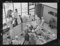
The other day someone showed a photo of an angry person with the caption of a neurosurgeon responding to the complaint of the neurological status of his patient. Since the days of the pioneers of neurosurgery including Harvey Cushing we as a specialty have always struggled to prove our relevance and necessity to society. Among our medical colleagues they often feel sympathy for our hard work but doubt that it really helps most of our patients. Similarly the public often views us at times heroically or other times seriously impotent to treat hopeless conditions.
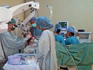
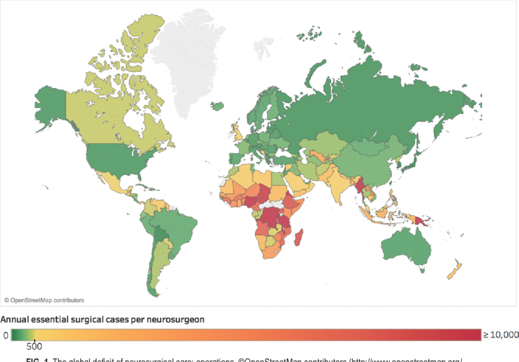
In 2018 a global survey of neurosurgery (see the enclosed photo)showed growing around the world and even in developing countries. This is a good thing. At the same time over the past several decades there has been increasing emphasis from stakeholders and political authorities for medical providers to show the effectiveness of their work and costs. What doctors do is now called relative value units and it is matched against assigned niches for our small specialty in health care budgets (both public and private).
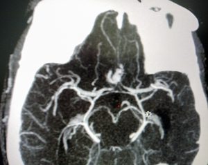
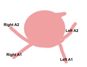
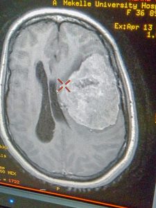
Since I was resident about 40 years ago wonderful developments have occurred such as MRI, intravascular intervention for aneurysm, spinal instrumentation, computerized navigation, endoscopic procedures etc. As a resident we are fascinated by these wonders and the science behind them but then when residency is over we do have to make our way in the real world. It is easy and totally understandable to be drawn to lucrative fields especially spine surgery as I was because it offers frankly good rewards for hard work as well as being so ubiquitous in the population. Unfortunately it is also the field most rift with poor science and loose surgical indications such that many of our medical colleagues tend to look down upon those whose career mostly deals with this field.
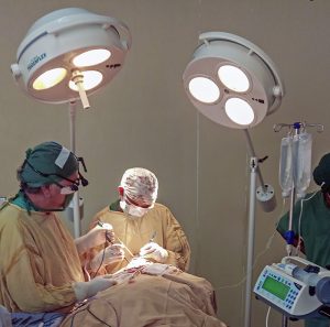
The enthusiasm for complicated cases of the brain and head injury treatment often wane as one passes from the academic training environment to the realm of sustaining oneself in the world which is understandable. In discussing this I am not passing any judgment just reflecting upon a fact of life.
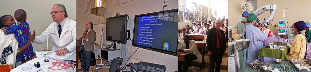
My advice to young neurosurgeons including my residents and fellows as well as especially those developing their careers in countries where neurosurgery is relatively new is to try to balance your contributions to society with your own financial rewards. Stay involved in teaching and research. Joining with public health officials in documenting the needs of traffic and accident victims will make society see the need for advancing neurosurgical care. Cooperate with fellow neurosurgeons and the medical community to educate stakeholders and public about how emergency care and neurosurgical centers (including trauma centers) can save so many who will continue to lead productive lives. Promote professional standards about spine care and be wary of becoming too entwined in the medical legal system. During surgery on patients who are not a risk of neurological deterioration but are likely to continue to be chronic pain sufferers does not improve your reputation or that of your profession. Let neurosurgery be the light of your medical community.

Neurosurgeon’s Day-Our Connection to the Harvey Cushing Legacy


Harvey Cushing-Founder and Innovator of Modern Neurosurgery

Very few times in the history of medicine has one man so dominated the development of a specialty that his birthday is celebrated as the day to recognize the field but that is case in Neurosurgery. The legacy of Harvey Cushing connects Ethiopian neurosurgery to his life’s work. When I entered Harvard Medical School in the Fall of 1977 I knew was interested in the field of neuroscience but I did not at first understand how I had entered into the hallowed historical grounds where Harvey Cushing made so many advances.

Harvard Medical School

Harvey was destined to be a fourth generation physician who began his studies at Harvard Medical School after undergrad at Yale just before the beginning of the 20th century. After graduation in 1895 he and Ernest Codman pioneered physiologically monitoring in anesthesia which dramatically reduced the previous death rates. Subsequently he trained in general surgery at Johns Hopkins under William Halsted, the great pioneer of modern general surgery. Residency training at Hopkins was scientifically driven by William Osler who also mentored Cushing leading him to write a biography of Osler in 1926 which won a Pulitzter prize.

From 1902 to 1937 first at Harvard, then Hopkins, and finally at Yale, Cushing performed over 2000 operations of the brain pioneering surgical techniques for the treatment of brain tumors. He revealed how the physiology of the brain functions in terms of blood pressure and brain perfusion and how the pituitary gland works. Brain surgery went from being a last chance high risk procedure to having the ability to safely save lives through out the world because of his influence and teaching.

Dr. Cushing performing surgery

As a medical student at Harvard I  saw patients on the grounds where Cushing lectured and went to operating room where Dr. Cushing performed brain surgery. I was fascinated and inspired by the life of Dr. Cushing and this experience in my early years led to my doing a special training in neuroscience as a medical student and then to train in neurosurgery after graduation.Later as resident in Neurosurgery at the University of Miami, Dr. Larry Page, who was trained at Harvard and the  Boston Children’s Hospital by neurosurgeons who trained under Cushing , was our main brain tumor training surgeon.

Six years ago I started the Neurosurgery program at Ayder Comprehensive Specialized Hospital-Mekelle University in Ethiopia where today we perform over 1000 operations a year. We currently are training 16 neurosurgeons for different regions in Ethiopia as well as Somaliland and Somalia. Following Cushing’s model of a neurosurgeon who cares about the world and his patients and uses the scientific model of research to improve life, we have created a multidisciplinary research team which has made significant discoveries in the epidemic of neural tube defects in Ethiopia. Our research led to the government studying a novel prevention program of fortifying salt with folic acid.


Professor Tony Magana performing surgery at Ayder Comprehensive Specialized Hospital-Mekelle University, Ethiopia

Happy Birthday to Harvey Cushing  you changed the world and helped inspire a kid from a small town in Texas to follow your example.


Advances in Subarachnoid Hemorrhage Treatment in Ethiopia

At Mekelle University Department of Neurosurgery-Ayder Comprehensive Specialized Hospital we have been developing an expertise in the treatment of stroke due to rupture of a cerebral blood vessel culminating in a successful clipping of a ruptured aneurysm.

Subarachnoid hemorrhage in Africa and Ethiopia
Stroke in Africa and more specifically in Ethiopia remains an almost taboo subject. It is shrouded in superstitious beliefs of curses and hidden poisons among most of the population who receive little public health education in what schooling they attend. A significant form of stroke is that due to rupture of a cerebral artery which creates the phenomena of subarachnoid hemorrhage. It is estimated that worldwide 9 in 100,000 years of human life or 1 in 50 people will suffer a subarachnoid hemorrhage.

Although rupture of a brain artery causing subarachnoid hemorrhage may lead to sudden death there are many patients who if given advanced treatment can be saved and return to functional lives. To receive this treatment requires special trained medical centers with experts in emergency medicine, neurology, radiology, anesthesiology, and neurosurgery. Up to now these centers have been lacking in most of Africa.

How subarachnoid hemorrhage causes damage
When a brain artery ruptures it may cause severe pressure on the brain which can kill or permanently disable. This type of large clot is unusual in most patients. Instead what happens is that the blood causes surrounding blood vessels to defensively constrict limiting the blood supply to the brain. This pathological process is vasospasm. Additionally the blood leakage can lead to chemical abnormalities of sodium or the mal-absorption of a fluid called cerebrospinal fluid which normal is produced and absorbed in a balanced way. Once a blood vessel ruptures once it will likely rupture again as each day goes by, a ticking time bomb.

Treatment of subarachnoid hemorrhage and ruptured cerebral aneurysms
Successful treatment of ruptured cerebral artery aneurysms requires rapidly making the diagnosis and beginning aggressive resuscitation of vasospasm and electrolyte abnormalities. The blood pressure must be closely controlled and the patients respiratory system supported. Upon stabilization the patient should undergo timely surgery or intravascular treatment to reduce the incidence of a second deadly rebleed. Whether microsurgery or intravascular treatment is better remains controversial.

A representative case at Ayder Comprehensive Specialized Hospital 
The following case is an example. A 55 year old Ethiopian grandmother suddenly complains of the worst headache of her life and goes into a coma. She is brought to Ayder Comprehensive Specialized Hospital in Mekelle, Ethiopia on the Mekelle University medical campus. Emergency physicians and internal medicine specialists stabilize her condition and perform a CT Scan which shows subarachnoid hemorrhage and suspician of a ruptured anterior communicating artery aneurysm.


A CT angiogram shows an anterior cerebral artery aneurysm

The patient is comatose with electrolyte abnormalities and out of control high blood pressure. She is admitted to the medical intensive care unit where she receives supplemental oxygen, high doses of fluids to correct hyponatremia and try to overcome the vasospasm, as well as a special medication, nimodipine, which can help to counteract vasospasm.

After 2 weeks she regains consciousness and a repeat CT angiogram ( a special CT scan which shows the arteries of the brain in detail ) is done which now clearly shows a 5mm aneurysm. Now that she is stable surgery must be done soon before a fatal rebleed can occur.

A large ballon is seen coming from a normal blood vessel which gets larger and thinner with time eventually rupturing

She is taken to the operating room with a specially trained anesthesia team which finely controls her blood pressure during surgery. An opening is made in the front and side of the skull while under general anesthesia and carefully working under the brain the ruptured blood vessel is exposed and clipped to prevent rebleeding.

Skull xray shows a clip has been placed closing the rupture aneurysm
The large aneurysm has been clipped while preserving the normal flow of blood to the brain


Should Surgeons other than Neurosurgeons Treat Hydrocephalus in Africa?

An illustration of a ventricular peritoneal shunt

Traditionally surgery for hydrocephalus has been performed by neurosurgeons who undergo vigorous neuroscience basic training and supervised surgical experience of at least 5 years in length (longer in developed countries) after medical school. Recently there has been some discussion as to whether in African and other undeveloped countries general surgeons should be taught to perform ventriculoperitoneal shunts to treat hydrocephalus. The treatment of hydrocephalus should remain under the direction and in the hands of neurosurgeons.

Hydrocephalus as a medical condition has been recognized since the time of the ancient Greeks. The concept of surgery to treat hydrocephalus by diverting the flow of cerebrospinal fluid began in  1949, when Nulsen and Spitz implanted a shunt successfully into the caval vein with a ball valve. Between 1955 and 1960, four independent groups invented distal slit, proximal slit, and diaphragm valves almost simultaneously.

An estimated 750,000 people have hydrocephalus, and 160,000 ventricular peritoneal shunts are implanted each year worldwide almost always by neurosurgeons. About 56,600 children and adolescents younger than age 18 years have a shunt in place.

The incidence of hydrocephalus in Africa is estimated to be 145 per 100,000 which is three times higher than in the developed world. Thousands of these children will need surgical intervention, either ventricular peritoneal shunt or the newer, but still not clearly accepted as superior, endoscopic procedures. 

A survey conducted among African neurosurgeons in 1998 showed that there were 500 neurosurgeons in Africa; that is, one neurosurgeon for 1,350,000 inhabitants, and 70,000 km2. That number is significantly increased now but the exact current number is unknown. Worldwide the average is 1 neurosurgeon per 230,000 but in Africa it can be as low 1 per 9 million people. It is believed there are 700 neurosurgeons currently or about 1 per 1,238,000  people which is an improvement but still not nearly enough. Ethiopia currently has about 30 practicing neurosurgeons and will soon be graduating about 30 newly trained neurosurgeons per year. This means Ethiopia will need about 450 functioning neurosurgeons taking into account expected population growth. 

Although the training of neurosurgeons in performing ventriculoperitoneal shunts has become somewhat standardized via the World Federation of Neurosurgical Societies as well as international neurosurgical groups, the training of general surgeons to do this procedure has not been rigorously studied. There are very few publications about the results of general surgeons performing ventriculoperitoneal shunts but an a study from Kenya done in 2010 showed a significantly high complication rate of 65% with an infection rate of 9.1% and shunt malfunction rate of 11.1%. This was much different than reported by Dr. Warf , an American trained neurosurgeon who created a specialized center in Uganda, with a malfunction rate of 4%.

More recently endoscopic procedure to open the third ventricle to the cistern and coagulate the choroid plexus are gaining ground but not totally proven yet. These procedures clearly require specialized training and knowledge of anatomy of the caliber of a neurosurgeon and not a general surgeon.

The real issue was not really the shortage of surgeons but the bottleneck was lack of hospitals, operating rooms, and clinics. Additionally transportation to get healthcare is a real issue. Now Ethiopia has seen the light and has  three training programs for neurosurgery in Ethiopia and are graduating about 30 per year. Again the problem is we have more surgeons than facilities to operate in.

African governments will see the idea of adding shunt placement to general surgery as an easy fix. In reality they are already overworked. I have taught medical students and general surgery residents. Many people think ventriculoperitoneal shunts are the easiest procedure but I always tell my neurosurgery residents and fellows it is not. Decisions about when to shunt, is the shunt working, is it infected? require experience and training. Academic following of outcomes, techniques, epidemiology requires an academic neurosurgery program take the lead.

Unfortunately there is no shortcut to capacity building. We are now training neurosurgeons for other African countries as well as Ethiopia. Eventually we will need more than 450 which will take time.

Finally I would say that our approach to hydrocephalus is changing rapidly. For us and the Uganda group we are consistently reducing the number of shunts we are doing each year. Whereas in the past we did nearly two hundred it is now going to be less than 100 even though we cover 20 million plus population with the highest myelomeningocoel rate in the world. We now recognize that many neural tube defect newborns have low grade infections which require antibiotics sometimes over 21 days until the csf is clear. Many times their hydrocephalus stabilizes after a few fontanelle taps. Although the post infectious group is rising ( we are in the infamous meningitis belt of Africa) similar we have avoided shunting by similar close follow-up. Our shunt infection rate is currently 3% because we identify these chronic low grade infections. I shutter to think what would happen if general surgeons with little experience are let loose upon this situation.


Finely Controlled Hypotension during Brain Surgery in Ethiopia

Brain surgery being done with controlled hypotension at Ayder Comprehensive Specialized Hospital

At Ayder Comprehensive Specialized Hospital, the university medical center for Mekelle University in Ethiopia, our experience with finely controlled hypotension during brain surgery for both adults and children has reduced the need for blood transfusion by half. 

In many underdeveloped African countries the surgical treatment of brain tumors is often very late in the course of the disease due to delay in the patient seeking treatment, having a diagnostic study to find the tumor, and being scheduled for surgery as many university centers have long waiting lists. Such is the situation we are in Ethiopia. These large brain tumors, often 10 centimeters or more in diameter, can require massive transfusion during the surgery to remove or reduce them. 

Large meningioma which has risk for high blood loss during surgery

The Department of Neurosurgery in the School of Medicine at Mekelle University in a close partnership with our Department of Anesthesia has been working on creating sustainable safe controlled hypotension techniques to reduce our blood loss during brain tumor surgery in adults and children at Ayder Comprehensive Specialized Hospital.  Thanks to the donation of a high quality intravenous perfuser by a diaspora American anesthesiologist and the cooperation of the university to gain stocks of Isoflurane  inhalation agent and Propofol intravenous agent as well as in house training together we have significantly reduced blood loss leading to much less transfusion during brain surgery. End tidal CO2 is kept at 4.5 to 5% and mean arterial blood pressure maintained at 65-70 mm/Hg.

brain surgery under controlled hypotension