What I learned and hope to return to at Mekelle University Ayder Comprehensive Specialized Hospital

Ayder Comprehensive Specialized Hospital campus of Mekelle University

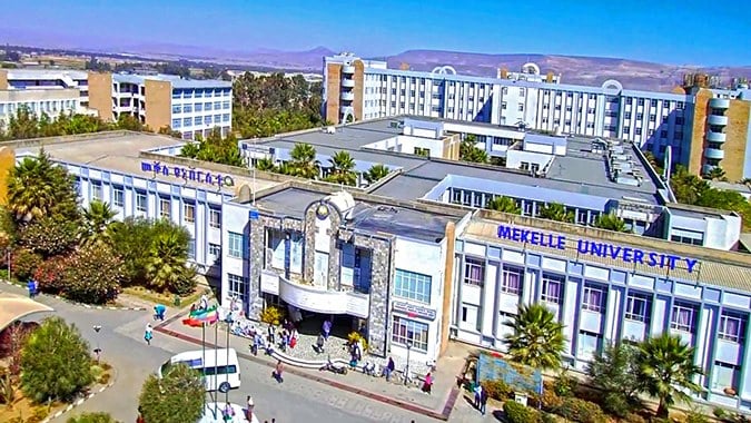
In building neuroscience and neurosurgery at Mekelle University I experienced great happiness in serving the people and training future neurosurgeons and scientists.  My seven year experience at Mekelle University serving the people of Tigray and surrounding areas as well teaching neurosurgeons and neuroscience gave me a new perspective on what is true career success. I now recognize there are three phases.

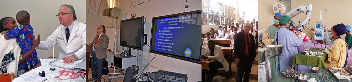
When I first came to Ayder Comprehensive Specialized Hospital in 2015 my initial goal was get a good neurosurgery service going. They were really the only government hospital capable of developing this goal outside Addis Ababa, the capital of Ethiopia, as well as being some distance north so it would offer chances to treat patients who would otherwise go untreated. The University and hospital were committed to building not only quality and quantity of good health care for Tigray and surrounding regions but also to medical education. 

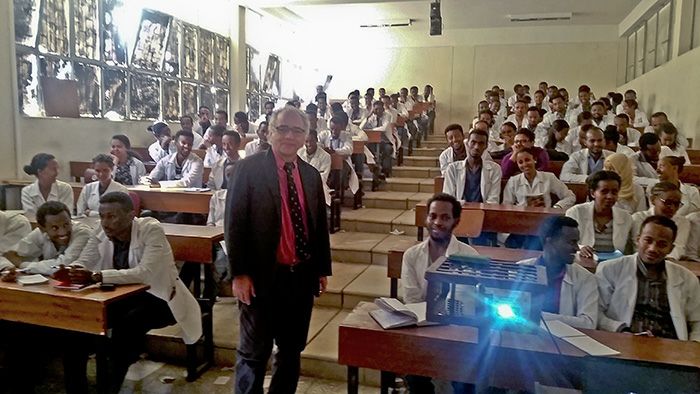
Initially I helped teach neuroscience and clinical neurology/neurosurgery to medical students and general surgery residents (young physicians who have graduated from medical school now doing specialty training) basic neurosurgical skills involving mostly traumatic injuries. My goal was to create a neurosurgery training program as well as a neuroscience research team. 

Teaching medical students about head injury

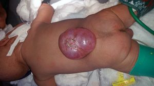
After about a year working with Ethiopian Ministries and some good collaboration from the Ethiopian ministries, World Federation of Neurosurgical Societies, and Mekelle University faculty we started a five year training program and a three year fellowship program. Up until the Tigray Ethiopian conflict we had grown to 18 trainees, doing 1,500 operations  year, and published internationally recognized research with our multidisciplinary research team on neural tube defects which occur in a high incidence in Ethiopia.

Our research team meet with the World Health Organization

Going through this experience made me reflect on what has been important in my career. When I was a college student at Texas A&M, then a medical student at Harvard, and finally a resident at the University of Miami I was mostly focused on personal achievement and perhaps also personal recognition. Through out that experience and subsequent practice I saw my career as having two phases. Learning to be a neurosurgeon and then becoming a great neurosurgeon. However it was my experience at Mekelle University and Ayder Comprehensive Hospital that taught me there was still a greater accomplishment which was to train great neurosurgeons and neuroscientists. 

Senior neurosurgery resident is supervised to remove a brain tumor

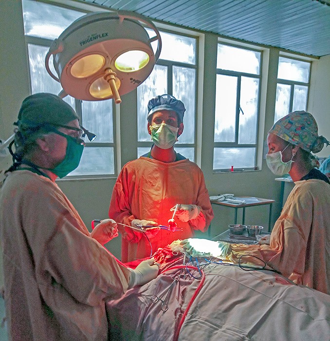
At first it was struggle. The hospital had little experience with neurosurgery. As a 60+ year old guy I was in the hospital every night doing surgery with young general surgeons and then residents with whom we had to start from scratch. Still they were eager to learn. With help from Indian and the few Ethiopian neurosurgeons we got up to date textbooks for them to read. Even though English was their second language they became proficient. At the end of the first year they knew more than I did at their level some 40 years ago. 

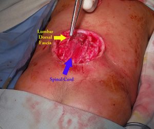
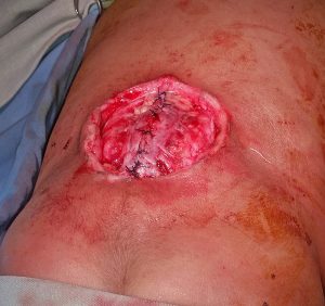
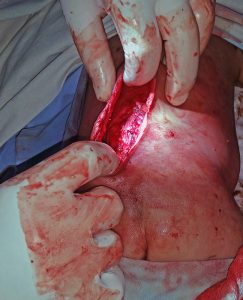
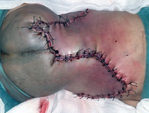
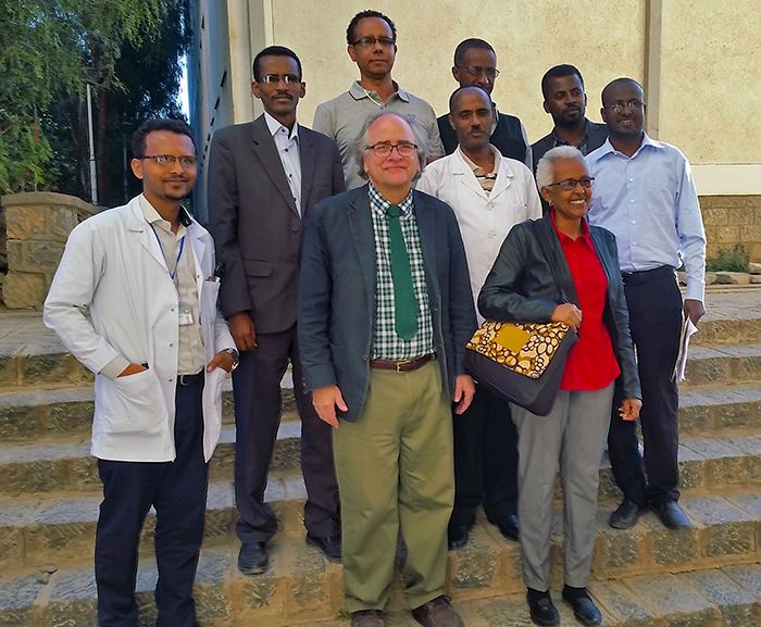
During surgery they have to learn about anesthesia, positioning, hand control, to make movements less than millimeter which could mean life or death, how to control bleeding, how to do a 12 hour operation, and much more. We began to have weekly seminars on very complicated ideas where they absorbed the concepts so well I was learning as much they were.  We were organized as most neurosurgical services in a sort of military style hierarchy to which the residents responded to well. Very quickly good and close relationships developed not only with those that were Tigray but also with others from other Ethiopian regions and other countries in the program.

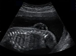
I formed strong relationships with the research bodies of Mekelle University and we created a strong multi-disciplinary research team on neural tube defects which lead to meeting with government and World Health Organization officials, international NGOs, and a finally the beginning of a nationwide plan for prevention and treatment.

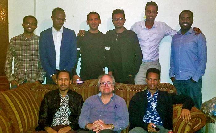
Before the  Ethiopian occupation and blockade shut the neurosurgery training program down we had operated on more than 5,000 patients from not only  Tigray but also Amhara,  Eritrea, and occasionally Addis Ababa to Gambella. This week far away from Mekelle I had been doing the “paper work” for my first graduates. I wish we could have had a formal ceremony but I had good voice communication provided. Now I have learned that the best phase of a neurosurgeon’s career is seeing his trainees carry on and expand what I started. 

Holiday dinner with my neurosurgery residents and fellow

I pray God will see fit to facilitate Ayder Comprehensive Hospital and myself to return  to its service, teaching, and research missions once again.

Recommendation for Valproic Acid Restricted Use in Ethiopia

prenatal ultrasound of myelomeningocoel
Prenatal ultrasound showing fetus with lumbar myelomeningocoel

Given the already highest incidence of neural tube defects measured globally present in Ethiopia it would seem exceedingly urgent to immediately call for the cessation of use of Valproic acid, which has been shown to cause neural tube defects,  in women of child bearing age in Ethiopia except for special circumstances.

History of Valproic Acid

Valproic acid was first produced in 1882. At that time its discoverer did not imagine that would be any therapeutic use because it was thought to have no pharmacological value. Then in the 1960s it was begun to be used in France and then in the late 1970s was approved for use in the United States as a primary drug for first epilepsy and then also for migraine headaches as well as mood disorders.

Over the next thirty years, however, concerns began to be raised about the potential relationship of the drug and birth defects.  Both a joint European study and a separate French study showed that women of child bearing age taking appropriate therapeutic doses of 1000 mg a day or more had a high incidence of neural tube defects and other malformations up to seven times higher than the control population.

Valproic Acid Use in Ethiopia

The use of Valproic acid is relatively new in Ethiopia in just the last few years. A review of the treatment of epilepsy from Gondar University showed that 4.84 % of patients were using it. In addition it is being used to treat mood disorders and migraine headache to an unknown extent. The prescription of this medicine is relatively unrestricted and can be done by general practitioners and even non-physician health care providers. Unfortunately the official formulary for Ethiopia briefly mentions that pregnancy is a contraindication and suggests consideration for folic acid supplementation should be given to reduce the risks of birth defects. However a recent animal model study using chick embryos showed that supplemental folic acid for neural tube defects caused by valproic acid was not effective in prevention.

Neural Tube Defects in Ethiopia

Our research at Mekelle University looking at the incidence of neural tube defects in the Tigray region of Northern Ethiopia suggested an over all incidence of at least 130 per 10,000 births but with some locals reporting close to 300 per 10,000 births. We know from studies done by the Ethiopian Institute of Public Health that 28% of women of children bearing age throughout Ethiopia have a significant folic acid deficiency which is likely the strongest contributing factor to the high incidence of neural tube defects

Recommendations for Valproic Acid Restriction in Ethiopia

1.Given the already highest incidence of neural tube defects measured globally present in Ethiopia it would seem exceedingly urgent to call for the immediate cessation of use of Valproic acid in women of child bearing age except for situations where no other drug can be used and the patient is receiving implantable or injectable forms of birth control.

2. Prescription of Valproic acid should only be done by physician sub-specialists in neurology, neurosurgery, or psychiatry and should require extensive counseling to the patient.

3.The formulary of Ethiopia should be modified and vigorously amended to warn of the risks. Although consideration for folic supplementation is appropriate it should be clearly stated that such supplementation may not be effective in preventing birth defects.


Food and Salt Fortification to Prevent Neural Tube Defects in Ethiopia

A joint study by the Departments of Neurosurgery and Nutrition (School of Public Health) of Mekelle University and the Department of Epidemiology of Emory University has concluded that thousands of newborn deaths and lifelong disability from neural tube defects could be prevented by fortifying food and possibly salt with folic acid to significantly reduce the epidemic currently occurring in Ethiopia.

“High potential for reducing folic acid‐preventable spina bifida and anencephaly, and related stillbirth and child mortality, in Ethiopia” is being published in Birth Defects Research.


New Recommended Action Plan for Prevention of Neural Tube Defects in Ethiopia

Over the past year in response to the Mekelle University Multidisciplinary Research team publications on neural tube defects in Tigray we have been interacting with the Ministry of Health, Maternal and Child, and the Ethiopian Public Health Institute. Two months ago Dr. Afework Mulugeta and myself as well as invited international experts gave a scientific advisory of this problem to the EPHI.

Yesterday a brief was given by the EPHI which was mostly based upon our research and recommendation as well as their review of available evidence.

Officially now the Ethiopian government recognizes “that there is an alarmingly high rate of neural tube defects and folate acid deficiency in Ethiopia”. The following recommendations were made to the higher ministry officials for approval
1. Periconceptional oral folate supplementation for all women of reproductive age as an immediate solution. Low cost imported folic acid is available for the public to purchase.
2. Making the first visit for pregnancy earlier instead of the current 16 weeks
3. Promoting the consumption of folic-rich foods.
4.Implementing mandatory wheat-flour food fortification.
5.Considering salt fortification with folic acid after doing a pilot study in Tigray
6.Establishing a surveillance system for NTDs throughout Ethiopia
7. Awareness creation for all recommended interventions strategies for the prevention of NTDs
Further studies will be done on community basis and on salt
Monitoring of food fortification measures to evaluate effectiveness

Difficulties in Preventing Neural Tube Defects in Ethiopia

Ethiopian mother hold her child with lumbar myelomeningocoel

The Mekelle University Multidisciplinary Research Group for Neural Tube Defects has just published its first research paper in Brain & Development Journal July 2018, “Maternal Risk Factors Associated with Neural Tube Defects in the Tigray Region of Ethiopia”. This being the first major prospective study done on neural tube defects in Ethiopia confirmed our worst fears of a very high incidence, significantly higher than the 75 per 10,000 births seen in most of Sub-Saharan Africa. There a many challenges in how this problem can be addressed involving cultural beliefs and practices, poverty, diet diversity, supplementation, and fortification.  Unfortunately, applying a Western style solution for Ethiopia will not be so easy or likely to succeed as well.

For the past decade the development of neurosurgery in Ethiopia has witnessed the high incidence of neural tube defects including myelomeningocoel and anencephaly coming to their clinics first in Addis Ababa but now also in Mekelle, Gondar, Bahir Dar, and Oromia. Previous published reports noted incidences first of greater than 160 per 10,000 births in Addis with a more recent report of 191 per 10,000 births in Addis Ababa. The reports of up 300 per 10,000 in some areas of Tigray are higher than those reported in any other developing countries.

Research replicated in nine countries in the 1960s and 1970s showed that neural tube defects were somewhat but no totally related to lack of folic acid in the diet and that adding folic acid would significantly reduce the incidence of neural tube defects. At first attempts were made with prescribing supplementation for women of childbearing age but this did not have the desired result. The incidence really came down in Europe and the United States when the government mandated the fortification of folic acid in food staples like bread and cereals

One of the factors we identified was that a lack of diet diversity increased the risk for NTDS while increased diversity reduced it. About 65% of the diet of most Ethiopians is enjera bread made at home from teff and boiled chick peas called shiro.   Although a serving of raw chick peas has about 1000 micrograms of folic acid its likely that boiling them reduces the folic acid to basically nothing.   A similar situation exists for spinach which is often boiled and eaten during the rainy season.

Traditionally, Ethiopians avoid eating fresh vegetables and fruits as documented in our study and many previous others. In fact a study of the one hundred most elite Ethiopian runners showed that 20% had a significant folic acid deficiency which correlated with lack of dietary diversity (avoiding greens and fruit). In the countryside where the women may spend many hours a day just to get 5 gallons of water, there is not much water to spare for cleaning produce. The population fears getting diarrheal illness from such produce.

Our study showed that of more than 13,000 women interviewed who were pregnant essentially none of them had any knowledge of preconceptional nutritional needs or about neural tube defects. 

Convincing Ethiopians to take medication for invisible illness such as hypertension has proved difficult. Research in many parts of Ethiopia has shown for example that as few as 50% of those prescribed medication for hypertension actually take it. There exists underlying fears of addiction to “un-natural” substances. Will Ethiopian women be convinced to take supplementation?

Ethiopians especially the 88% who live in country side rarely buy food staples like bread but instead make their own enjera from stored teff. Currently there is only one factory in the country capable of making fortified bread but it is not functioning.

The cost of a months supply of folic acid 4 milligrams per day is about 80 birr or about $2.40 US for a single woman. Given the fact that the average family makes about 250 birr per month to support a family of six, there is little room to allow payment of this expense. There are no current domestic producers of folic acid so considerable hard foreign currency would need to be mobilized to import stock. For the government to provide this for every women of child bearing age would cost tens of millions of dollars to be added to the budget of a country which currently spends the equivalent of about $13 per capita for the 100 million population.

Ultimately addressing the issue of the high incidence of neural tube defects in Ethiopia will be requiring taking into account cultural norms and practices in such a way that changes are seen as consistent with Ethiopian culture.  Widespread public education and a major investment in folic acid purchases by the government will be necessary. This begins with the clear realization that there is a problem.


Research:Neural Tube Defects in Tigray Ethiopia

Recent research we have done at Mekelle University soon to be published has confirmed that there is a high rate of neural tube defects affecting the brain and spinal cord in Tigray. Experience suggests this is also the case in other parts of Ethiopia as well. At least 131 out of every 10,000 births is affected with some areas having almost twice that number. Defects result in death at birth for about 77% of the pregnancies affected, usually with anencephaly, while the 23% born alive usually have severe paralysis of the lower extremities and often need a operations to close the open spine, closure of myelomeningocoel,  and to control pressure in the brain, ventricular peritoneal shunt, for lumbar and thoracic myelomeningocoel associated with an Arnold Chiari II malformation causing hydrocephalus. Lesser numbers of encephalocoel often requiring closure were encountered as well.

Ethiopian mother hold her child with lumbar myelomeningocoel

The most likely significant cause is lack of diversity in the diet and especially failing to consume foods with the vitamin folic acid. This is usually found in green and leafy vegetables as well as fresh fruits. Cooking foods such as chick peas which may contain folic acid will destroy much of it.

Around the world these birth defects have been reduced about 75% by encouraging women to plan their pregnancy and take 4 milligrams of folic acid daily starting before conception. We are working with our research group at Mekelle University to help the Tigray Regional Health Bureau and the Ethiopian Ministry of Health come up with programs to reduce these defects but this will take time.

In the meantime we encourage all women in Ethiopia to plan their pregnancy, starting folic acid supplementation before they conceive, and practice dietary diversity. They should try to wait one year between pregnancies, and understand that breast feeding increases their need for folic acid. Very young and older women are more susceptible to having children with these defects. Other factors may be involved besides folic acid deficiency but the good news is that folic acid supplementation will likely still reduce these defects.

Lumbar Myelomeningocoel Surgery in Northern Ethiopia

This a review of some of the principals I follow when performing myelomeningocoel surgery in young infants in Northern Ethiopia. More than a third of all the neurosurgery operations is made up by this pediatric neurosurgical condition at our hospital.

It is estimated by the World Health Organization that over 300,000 children suffer neural tube defects which results in improper formation of the spinal cord, spine, and brain leading to absence of bowel, bladder, and sexual function control, spinal deformity, reduced mental development, and paralysis. More common in underdeveloped countries then in developed countries, the occurrence has been mostly strongly related to preconception maternal folate deficiency and some genetic factors. Unfortunately in Ethiopia there are thousands of these children born every year, we see several a week in our clinic at Ayder Comprehensive Specialized Hospital at the Mekelle University College of Health Sciences in Northern Ethiopia.

Myelomeningocoel is a disorder where the neural tube which develops in the first month after conception from a flat plate of specialized tissue fails to fully close into a tube. This results in neural elements being exposed directly to the atmosphere or only a thin membranous layer. The lowest nerves below the defect often do not completely form leading to paralysis in the lower extremities and difficulties with the functions normally performed by the sacral nerve roots involving the bladder, rectum, and sexual organs.

Pre-Operative Assessment

Pre-operatively many of these children come to the hospital from long distances in poor nutritional condition. If they weigh less than 3 kilograms we delay surgery until they reach this weight because of problems with temperature control in the operative and immediate post-operative period. Children who have cerebrospinal fluid leaks, chronic bleeding from the neural placode, progressive macrocephaly, symptomatic hydrocephalus, symptomatic Arnold-Chiari symptoms such as weak cry, weak upper extremities or multiple pneumonias from aspiration, or meningitis are given priority. Because of the large number of cases and limited resources there is often a several month waiting period for those in stable condition with well epithelialized neural tube defects.

To undergo surgery they must be fit for general anesthesia with normal hemoglobin, kidney function, cardiovascular status and be free of active infection. Ultrasound evaluations of the head to assess for hydrocephalus and of the abdomen to assess the kidneys is routinely done.

They are kept nothing per oral (NPO) for several hours before surgery and started on pediatric maintenance intravenous fluid containing glucose during this time which is continued in the operating room and after until the child is breast-feeding normally.

We do not routinely prepare blood for the surgery unless the child has a low hemoglobin/hematocrit preoperatively. If so then a pre-operative transfusion will be given.

Surgery

The child is placed under general inhalation anesthesia using isofluorane rather then halothane is the most common anesthetic agent used in Ethiopia. We use a electric heater in the room, warm the saline irrigation used during the case, intravenous fluid heaters, and keep the child covered as much as possible to avoid hypothermia which can occur easily in our mountain environment.

Pre-operative ceftriazone at 50mg/kg is given for prophylaxis and continued for 48 hours post-operatively.  This is given not just to prevent wound infection but also because these children are at risk of developing post operative pneumonia from the co-existing Arnold-Chiari malformation also present.

myelom-closure1

The child is placed in prone position with an intravenous fluid bag under the chest and under the groin which leaves the abdomen free of pressure thus reducing venous bleeding during the surgery. In the picture is seen a child with a large membranous lumbar myelomeningocoel. The large size taking up most of the width of the back and the mostly membranous covering predicts that closure will be difficult.

myelom-closure2

The dissection is done under loupe magnification. The neural elements are dissected free of the epithelium, fat, scar tissue, and fibrous bands. In the picture above the spinal cord with attached neural placode and the lumbar dorsal fascia have been identified.

myelom-closure3

In many cases the lumbar dorsal fascia can be carefully dissected at the lateral extant of the wound and then reflected medially to be closed as a dural layer. In the picture above this “dural” layer is closed first by several interrupted 3-0 vicryl sutures and the oversewn with a running suture.

myelom-closure4

Key to being able to close the wound is mobilizing the subcutaneous fascia superiorly and inferiorly and bilaterally. This must be generously done by careful digital dissection to free up the skin edges. It is generally better to over mobilize then undermobilize to reduce tension on the skin closure. If the a primary midline closure cannot be done then the primary skin fascial incision can be extended to superior and inferior z-plasty incisions which must be longer than the original incision. Alternatively a rhomboid type flap can also be used. It is important in the post-operative period to support the flap by giving supplemental oxygen, maintaining hydration, checking the post-operative hemoglobin/hematocrit, and avoid undo pressure on the flap. Most of the cases can be primarily closed.

Despite the best techniques, about 20 percent of cases have some type of wound problem which can range from mild stitch abscess or dehisicience to severe dehisicience. I always tell the surgical residents that we close the dura thinking that should the flap come apart with a sound dural closure we have a good chance for secondary healing.

myelom-closure5

This was a very large myelomeningocoel that we attempted to close with a modified z-plasty.  Unfortunately a week after surgery there was some necrosis and breakdown. However, with aggressive daily wound care and good nutritional support a good result was obtained by secondary intention healing over a month’s time because of the good dural closure underneath.

Careful monitoring of the head circumference, anterior fontanel, eye movements, mental status, and feeding is necessary in the immediate and later post-operative period. Most of these children will go on to develop progressive hydrocephalus requiring a second operation for insertion of a ventricular peritoneal shunt. A few of them if they have persistent stridor, weak cry, aspiration will need to go undergo an upper cervical laminectomy and decompression of the brainstem for treatment of the Arnold-Chiari malformation.