Advances in Subarachnoid Hemorrhage Treatment in Ethiopia

At Mekelle University Department of Neurosurgery-Ayder Comprehensive Specialized Hospital we have been developing an expertise in the treatment of stroke due to rupture of a cerebral blood vessel culminating in a successful clipping of a ruptured aneurysm.

Subarachnoid hemorrhage in Africa and Ethiopia
Stroke in Africa and more specifically in Ethiopia remains an almost taboo subject. It is shrouded in superstitious beliefs of curses and hidden poisons among most of the population who receive little public health education in what schooling they attend. A significant form of stroke is that due to rupture of a cerebral artery which creates the phenomena of subarachnoid hemorrhage. It is estimated that worldwide 9 in 100,000 years of human life or 1 in 50 people will suffer a subarachnoid hemorrhage.

Although rupture of a brain artery causing subarachnoid hemorrhage may lead to sudden death there are many patients who if given advanced treatment can be saved and return to functional lives. To receive this treatment requires special trained medical centers with experts in emergency medicine, neurology, radiology, anesthesiology, and neurosurgery. Up to now these centers have been lacking in most of Africa.

How subarachnoid hemorrhage causes damage
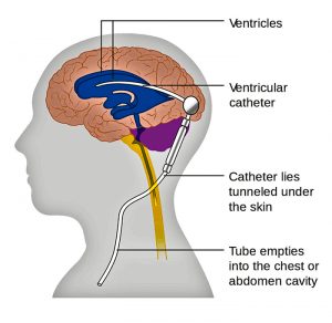
When a brain artery ruptures it may cause severe pressure on the brain which can kill or permanently disable. This type of large clot is unusual in most patients. Instead what happens is that the blood causes surrounding blood vessels to defensively constrict limiting the blood supply to the brain. This pathological process is vasospasm. Additionally the blood leakage can lead to chemical abnormalities of sodium or the mal-absorption of a fluid called cerebrospinal fluid which normal is produced and absorbed in a balanced way. Once a blood vessel ruptures once it will likely rupture again as each day goes by, a ticking time bomb.

Treatment of subarachnoid hemorrhage and ruptured cerebral aneurysms
Successful treatment of ruptured cerebral artery aneurysms requires rapidly making the diagnosis and beginning aggressive resuscitation of vasospasm and electrolyte abnormalities. The blood pressure must be closely controlled and the patients respiratory system supported. Upon stabilization the patient should undergo timely surgery or intravascular treatment to reduce the incidence of a second deadly rebleed. Whether microsurgery or intravascular treatment is better remains controversial.

A representative case at Ayder Comprehensive Specialized Hospital 
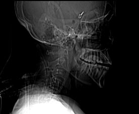
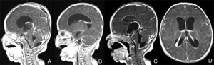
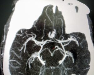
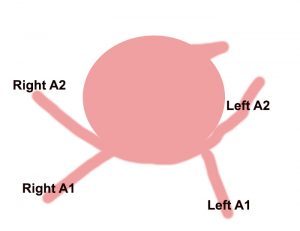
The following case is an example. A 55 year old Ethiopian grandmother suddenly complains of the worst headache of her life and goes into a coma. She is brought to Ayder Comprehensive Specialized Hospital in Mekelle, Ethiopia on the Mekelle University medical campus. Emergency physicians and internal medicine specialists stabilize her condition and perform a CT Scan which shows subarachnoid hemorrhage and suspician of a ruptured anterior communicating artery aneurysm.


A CT angiogram shows an anterior cerebral artery aneurysm

The patient is comatose with electrolyte abnormalities and out of control high blood pressure. She is admitted to the medical intensive care unit where she receives supplemental oxygen, high doses of fluids to correct hyponatremia and try to overcome the vasospasm, as well as a special medication, nimodipine, which can help to counteract vasospasm.

After 2 weeks she regains consciousness and a repeat CT angiogram ( a special CT scan which shows the arteries of the brain in detail ) is done which now clearly shows a 5mm aneurysm. Now that she is stable surgery must be done soon before a fatal rebleed can occur.

A large ballon is seen coming from a normal blood vessel which gets larger and thinner with time eventually rupturing

She is taken to the operating room with a specially trained anesthesia team which finely controls her blood pressure during surgery. An opening is made in the front and side of the skull while under general anesthesia and carefully working under the brain the ruptured blood vessel is exposed and clipped to prevent rebleeding.

Skull xray shows a clip has been placed closing the rupture aneurysm
The large aneurysm has been clipped while preserving the normal flow of blood to the brain


The Growing Infant Meningitis Crisis in Ethiopia


MRI of infant with meningits
Infant with meningitis seen on MRI to have hydrocephalus and meningeal enhancement

Ethiopia is facing a growing crisis in infant meningitis compounded by a false reliance on the power of antibiotics to treat the problem without definitive diagnosis and follow up.

A growing problem
As you may know north eastern Africa including Ethiopia is part of the so-called “meningitis belt” with the highest rates of bacterial meningitis in the world. Just a couple a months ago I was attending a meeting with Ministry of Health with the new five pediatric neurosurgery centers of excellence in Ethiopia where a new growing epidemic of chronic meningitis seen throughout the country was discussed. Over the past year we are seeing more and more cases of infants on an almost daily basis presenting with progressive hydrocephalus who have diagnostic cerebrospinal fluid consistent with chronic meningitis. Our average census is 5 to 8 in hospital all the time whereas a few years ago it was only one or two. Many of these children have hospitalizations of 12 weeks or more. Many have died and many are left with significant cerebral disability which could have been prevented by earlier intervention and appropriate treatment lengths.

Systemic deficiencies
While this epidemic seems to be worsening, Dr. Abreha (Head of Pediatrics and Pediatric Neurologist at Mekelle University) and myself are concerned that deficiencies in facilities and procedures within the medical system may be contributing to it. Unfortunately throughout Ethiopia almost no lumbar punctures are now done to diagnose meningitis. A sample inquiry of pediatric residents and interns found that 75% of them had never done a lumbar puncture. Additionally many clinics and hospitals do not even have lumbar puncture trays. A false sense of security exists that powerful antibiotics given a short period time can will solve the problem

Secondly, the lack of ability to process or even receive an CSF specimen for analysis beyond 5 PM forces the treating physicians to start treatment without first obtaining a culture which is against world wide standard of culture first then start treatment. This seems very contrarian to the frequent theme of the pharmacy and laboratory professionals in Ethiopia that there is abuse of antibiotics.

A key analysis is determining the glucose, protein, and cell count in cerebrospinal fluid. Dr. Abreha recently gave a sample of tap water to the laboratory to be tested on the automated machine and it diagnosed multiple WBC consistent with meningitis. The equipment supplied to Ethiopian hospital is not capable of accurate diagnosis. Technicians can do manual counts but these are time consuming and labor intensive. This inaccuracy has greatly impaired our ability to determine if patients are infection free which they must be before they can undergo definitive treatment for hydrocephalus which is a ventriculoperitoneal shunt.

Thirdly, around the world there is much controversy about for how long antibiotic treatment should be given ranging from 10 days to 21 days. The problem is that partially treated meningitis patients who have not been cleared from the infection often do not have fever, meningeal signs, or other clinical findings except hydrocephalus. Many infants are briefly seen for nonspecific fever and receive short courses of antibiotics in Ethiopia without specific diagnosis being made and without adequate follow up. Many of these children we believe are harboring these low grade chronic infections leading to their late appearance at Ayder  Comprehensive Specialized Hospital. This creates a great dilemma as these children often require treatment of intravenous powerful expensive antibiotics from 21 days to in excess of six weeks or more until the infection is cleared by the demonstration of two negative cultures off antibiotics and normal cell counts. In addition in order for the shunt to work the protein has to be less than 150 mg/dl. Failure to diagnose an active residual infection before a shunt placement will only aggravate the infection leading to shunt removal and complications.

The treasure and future of Ethiopia is in her children. Meningitis appropriately treated early and followed can have a very low morbidity and disability outcome. The current situation if not acted upon will result in increasing medical costs, increasing disability, and increasing infant death which could be prevented with simple directed community and institutional action. 

Recommended Course of Action
1. Immediate upgrade of laboratory ability in hospitals to receive CSF specimens 24 hours a day for culture, gram stain, sensitivity.
2. Training of interns and residents in performing lumbar puncture and making sets available
3. Institute an Ethiopian wide policy of obtaining CSF before starting antibiotics. Children with seizures, lethargy, focal deficit, or signs of increased intracranial pressure would be emergently sent to a referral hospital for CT Scan or if possible if they have an  open fontanelle undergo an head ultrasound locally to rule out mass before lumbar puncture.
4. Make sure all children treated for meningitis undergo follow-up and that a repeat lumbar puncture is done after treatment. This may seem over kill but given the crisis happening it is the only way to prevent these chronic cases.
5. Funding of community service, public education, and research projects on this vital issue in cooperation with the regional health bureaus.
6. Training of health officers, nurses, and general practitioners in proper diagnosis, evaluation, treatment, and referral of children with meningitis and hydrocephalus.

Should Surgeons other than Neurosurgeons Treat Hydrocephalus in Africa?

An illustration of a ventricular peritoneal shunt

Traditionally surgery for hydrocephalus has been performed by neurosurgeons who undergo vigorous neuroscience basic training and supervised surgical experience of at least 5 years in length (longer in developed countries) after medical school. Recently there has been some discussion as to whether in African and other undeveloped countries general surgeons should be taught to perform ventriculoperitoneal shunts to treat hydrocephalus. The treatment of hydrocephalus should remain under the direction and in the hands of neurosurgeons.

Hydrocephalus as a medical condition has been recognized since the time of the ancient Greeks. The concept of surgery to treat hydrocephalus by diverting the flow of cerebrospinal fluid began in  1949, when Nulsen and Spitz implanted a shunt successfully into the caval vein with a ball valve. Between 1955 and 1960, four independent groups invented distal slit, proximal slit, and diaphragm valves almost simultaneously.

An estimated 750,000 people have hydrocephalus, and 160,000 ventricular peritoneal shunts are implanted each year worldwide almost always by neurosurgeons. About 56,600 children and adolescents younger than age 18 years have a shunt in place.

The incidence of hydrocephalus in Africa is estimated to be 145 per 100,000 which is three times higher than in the developed world. Thousands of these children will need surgical intervention, either ventricular peritoneal shunt or the newer, but still not clearly accepted as superior, endoscopic procedures. 

A survey conducted among African neurosurgeons in 1998 showed that there were 500 neurosurgeons in Africa; that is, one neurosurgeon for 1,350,000 inhabitants, and 70,000 km2. That number is significantly increased now but the exact current number is unknown. Worldwide the average is 1 neurosurgeon per 230,000 but in Africa it can be as low 1 per 9 million people. It is believed there are 700 neurosurgeons currently or about 1 per 1,238,000  people which is an improvement but still not nearly enough. Ethiopia currently has about 30 practicing neurosurgeons and will soon be graduating about 30 newly trained neurosurgeons per year. This means Ethiopia will need about 450 functioning neurosurgeons taking into account expected population growth. 

Although the training of neurosurgeons in performing ventriculoperitoneal shunts has become somewhat standardized via the World Federation of Neurosurgical Societies as well as international neurosurgical groups, the training of general surgeons to do this procedure has not been rigorously studied. There are very few publications about the results of general surgeons performing ventriculoperitoneal shunts but an a study from Kenya done in 2010 showed a significantly high complication rate of 65% with an infection rate of 9.1% and shunt malfunction rate of 11.1%. This was much different than reported by Dr. Warf , an American trained neurosurgeon who created a specialized center in Uganda, with a malfunction rate of 4%.

More recently endoscopic procedure to open the third ventricle to the cistern and coagulate the choroid plexus are gaining ground but not totally proven yet. These procedures clearly require specialized training and knowledge of anatomy of the caliber of a neurosurgeon and not a general surgeon.

The real issue was not really the shortage of surgeons but the bottleneck was lack of hospitals, operating rooms, and clinics. Additionally transportation to get healthcare is a real issue. Now Ethiopia has seen the light and has  three training programs for neurosurgery in Ethiopia and are graduating about 30 per year. Again the problem is we have more surgeons than facilities to operate in.

African governments will see the idea of adding shunt placement to general surgery as an easy fix. In reality they are already overworked. I have taught medical students and general surgery residents. Many people think ventriculoperitoneal shunts are the easiest procedure but I always tell my neurosurgery residents and fellows it is not. Decisions about when to shunt, is the shunt working, is it infected? require experience and training. Academic following of outcomes, techniques, epidemiology requires an academic neurosurgery program take the lead.

Unfortunately there is no shortcut to capacity building. We are now training neurosurgeons for other African countries as well as Ethiopia. Eventually we will need more than 450 which will take time.

Finally I would say that our approach to hydrocephalus is changing rapidly. For us and the Uganda group we are consistently reducing the number of shunts we are doing each year. Whereas in the past we did nearly two hundred it is now going to be less than 100 even though we cover 20 million plus population with the highest myelomeningocoel rate in the world. We now recognize that many neural tube defect newborns have low grade infections which require antibiotics sometimes over 21 days until the csf is clear. Many times their hydrocephalus stabilizes after a few fontanelle taps. Although the post infectious group is rising ( we are in the infamous meningitis belt of Africa) similar we have avoided shunting by similar close follow-up. Our shunt infection rate is currently 3% because we identify these chronic low grade infections. I shutter to think what would happen if general surgeons with little experience are let loose upon this situation.


Alternative Ventricular Peritoneal Shunt Anchoring Method in Pediatric Patients with Chhabra Shunt System

Object: The author reports his experience using the Chhaabra Slit n Spring shunt system SH202 in pediatric patients in Ethiopia using connectors at the entry point of the shunt into the ventricle to anchor the system without anchoring suture. Placing an anchoring suture is often difficult in very young pediatric patients because of a lack of available tissue. This alternative was therefore developed.

Presented at the 21st Meeting of the Surgical Society of Ethiopia at the African Union, Addis Ababa, Ethiopia on February 3, 2017.

Methods: The consecutive results of patients was retrospectively studied from 10/7/2014 to 9/21/2016 in 76 patients. Follow-up ranged from 26 months to 3 months through December 2016. Most the patients were 6 months of age or less (49) while (16) were between 6 to 12 months and (11) were greater than 1 year of age.

The ventricular catheter was connected to a straight connector which was connected to silicone tube which bent at the entry into the brain. The silicon tube proximal end was connected via connector the shunt valve. All connections were secured with 2-0 silk. Otherwise the procedure is as described by B.C. Warf in J Neurosurg (Pediatrics 4) 102:358–362, 2005. Complications were 7% shunt revision , 4% minor wound revision, and 5% shunt infection.

Conclusion:This alternative method of anchoring the shunt without suture compares favorably with other previous reports of the Chaabra shunt system. There were no intraventricular migrations or migrations out of the ventricle as has been reported in the literature. When revision was necessary it was slightly more complicated than with the ventricle single connector setup but still could be accomplished without too much effort. Longer follow-up is necessary to more fully evaluate this alternative.

PDF of slide presentation

sse2017-shunt-proftony

Emergency Percutaneous External Ventricular Drainage for Infants

For the past 18 months we have used a novel technique for emergency percutaneous external ventricular drainage (EPEVD) in young infants. This is taught to pediatric residents and general surgery residents under the direction of the neurosurgical unit at Ayder Comprehensive Specialized Hospital, Mekelle University College of Health Sciences. Acute obstructive or communicating hydrocephalus presenting in young infants with patent anterior fontanels is a common occurrence in teaching hospitals of developing countries. This life saving procedure can allow a rapid decompression of intracranial pressure and the immediate safe acquisition of cerebrospinal fluid analysis for diagnosis.

The following case is an example of the procedure. A 6 month old child was seen by the parents to have progressive head growth and lack of development over several months. Examination showed a tense fontanel, macrocephaly, and developmental delay. A cranial ultrasound showed severe hydrocephalus with viscous intraventricular content consistent with infection.

We suspected tuberculosis of the ventricles and urgent decompression and diagnosis was necessary.

6 month old child with hydrocephalus due to chronic tuberculosis of the lateral ventricles

With the mother holding the child so no sedation or anesthesia was necessary, the scalp was prepped with betadiene. Then an 18 gauge angiocatheter was placed perpendicular to the mid-pupillary line  in the anterior fontanel.

Perpendicular placement of angiocath into lateral ventricle on mid-pupillary line in lateral anterior fontanel

The needle stylet is removed and the angiocatheter seated to scalp. Several gauze are used to wrap the angiocathether which will then hold it firm when a protective dome is placed.

The drainage system is connected and the catheter wrapped with sterile gauze to prevent movement laterally

The protective dome made out of the top of common water bottle that has been cleaned with alcohol is slipped over the drainage tube. By securing the catheter to prevent dislodgement or lateral movement the protective dome prevent secondary brain injury and displacement of the catheter.

The protective dome is placed over the catheter wrapped in gauze

The drainage tube and dome are then secured with tape to the skull. In this case because of the high viscosity of the intraventricular fluid only a small amount of elevation of the drainage tube over the head was used.

Final dressing of ventricular catheter drainage system

This is a useful temporary measure which does not require an operating room or advanced skill of a neurosurgeon to perform.  It does have the disadvantage of not being tunneled but is not intended for long term use. The use of easily available material in the setting of a developing country and teaching it to pediatricians and general surgeons in a country setting where few neurosurgeons are present will allow access of emergency treatment for more infants.