
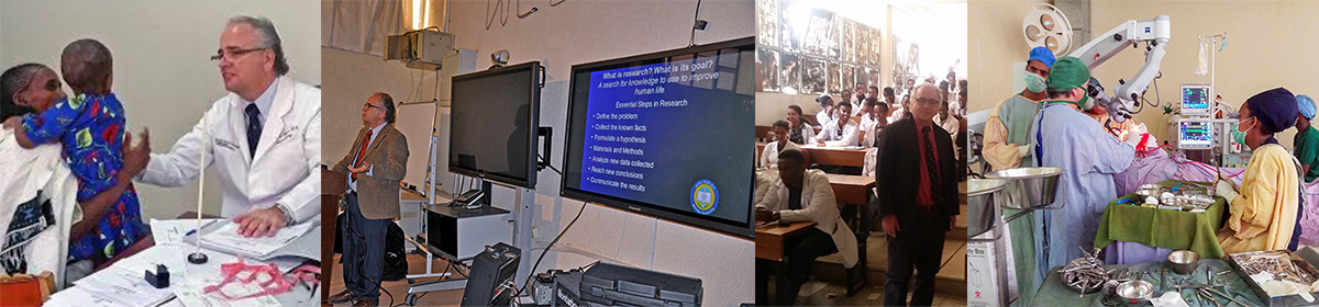
For the past 18 months we have used a novel technique for emergency percutaneous external ventricular drainage (EPEVD) in young infants. This is taught to pediatric residents and general surgery residents under the direction of the neurosurgical unit at Ayder Comprehensive Specialized Hospital, Mekelle University College of Health Sciences. Acute obstructive or communicating hydrocephalus presenting in young infants with patent anterior fontanels is a common occurrence in teaching hospitals of developing countries. This life saving procedure can allow a rapid decompression of intracranial pressure and the immediate safe acquisition of cerebrospinal fluid analysis for diagnosis.
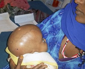
The following case is an example of the procedure. A 6 month old child was seen by the parents to have progressive head growth and lack of development over several months. Examination showed a tense fontanel, macrocephaly, and developmental delay. A cranial ultrasound showed severe hydrocephalus with viscous intraventricular content consistent with infection.
We suspected tuberculosis of the ventricles and urgent decompression and diagnosis was necessary.
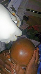
With the mother holding the child so no sedation or anesthesia was necessary, the scalp was prepped with betadiene. Then an 18 gauge angiocatheter was placed perpendicular to the mid-pupillary line in the anterior fontanel.
The needle stylet is removed and the angiocatheter seated to scalp. Several gauze are used to wrap the angiocathether which will then hold it firm when a protective dome is placed.
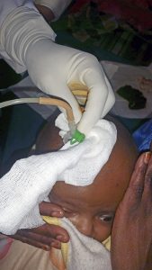
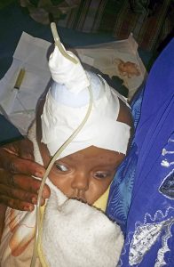
The protective dome made out of the top of common water bottle that has been cleaned with alcohol is slipped over the drainage tube. By securing the catheter to prevent dislodgement or lateral movement the protective dome prevent secondary brain injury and displacement of the catheter.
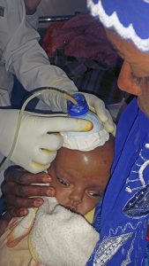
The drainage tube and dome are then secured with tape to the skull. In this case because of the high viscosity of the intraventricular fluid only a small amount of elevation of the drainage tube over the head was used.
This is a useful temporary measure which does not require an operating room or advanced skill of a neurosurgeon to perform. It does have the disadvantage of not being tunneled but is not intended for long term use. The use of easily available material in the setting of a developing country and teaching it to pediatricians and general surgeons in a country setting where few neurosurgeons are present will allow access of emergency treatment for more infants.